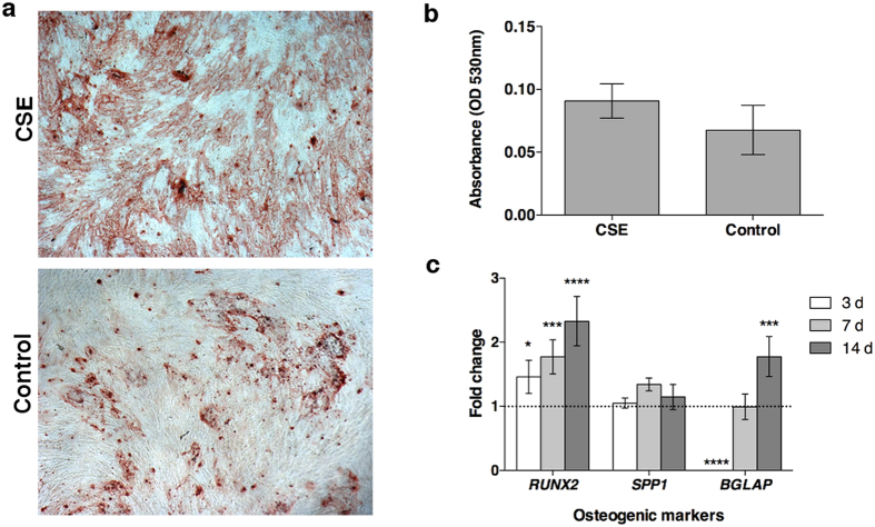
Figure 5. Calcium deposition and gene expression for osteogenic differentiation.
AdMSCs were seeded and allowed to attach overnight before being exposed to 0.5% CSE at the time of osteogenic induction. The CSE was allowed to remain on the cells for 24 h. (A) After 21 d in culture, the cells were stained for calcium deposition with Alizarin Red S solution (4× magnification). (B) The staining solution was removed and the absorbance was read at 530 nm. There were no significant differences detected from the staining after acute CSE exposure. (C) To examine the effects on gene expression an early transcription factor (RUNX2), an early structural protein (SPP1), and a late osteoblastic marker (BGLAP) were evaluated. At day 3, the differentiation seems to be slowed in cells exposed to CSE, however expression of late osteoblastic markers was increased after CSE exposure. (*p < 0.05, ***p < 0.001, ****p < 0.0001).

