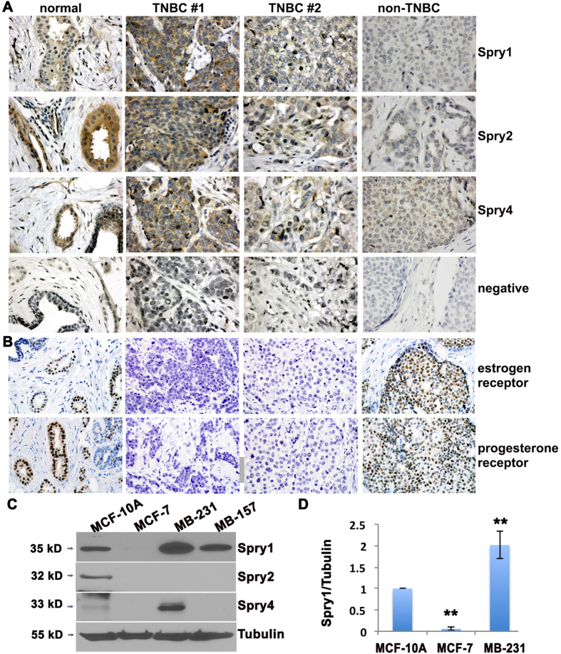
Figure 1. Spry1 is expressed in TNBC and in MDA-MB-231 TNBC cells.
(A) Representative views of normal breast tissue versus biopsy from non-TNBC and TNBC tissues immunostained to detect Spry1, Spry2 and Spry4 (brown reaction product). Tissues were counterstained with hematoxylin. For negative controls (NEG), sections were incubated with normal rabbit IgG instead of specific antibody. (B) Sections were stained to detect the estrogen receptor or progesterone receptor, and counterstained with hematoxylin. (C) Immunoblotting to evaluate the expression of Spry proteins in the TNBC cell line MDA-MB-231 and MDA-MB-157 compared to the normal mammary epithelial cell line MCF-10A and the non-TNBC cell line MCF-7. The blots were stripped off and reused for probing of Tubulin. (D) Quantification of Spry1 protein levels normalized by tubulin levels from three independent experiments. **p < 0.05 relative to controls. Images are from original 400x magnification.

