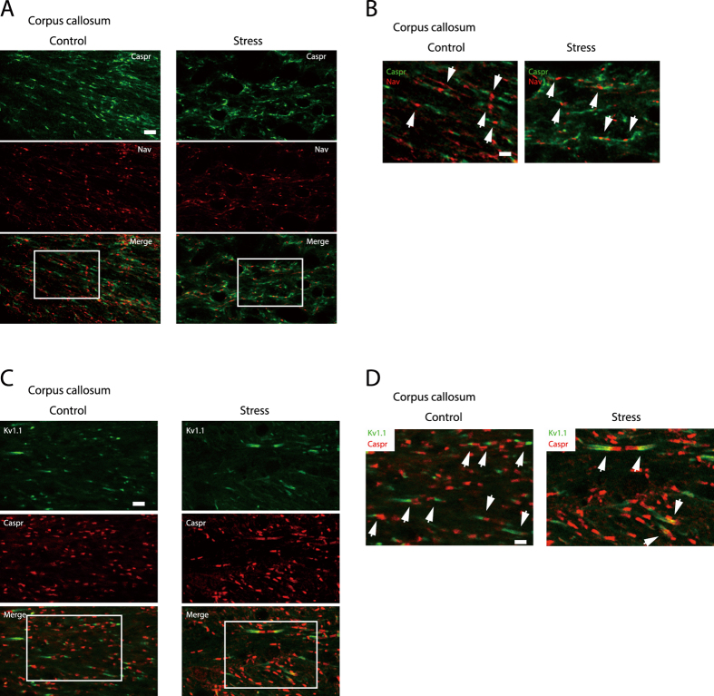
Figure 2. Increase in the length of Caspr- and K v1.1-immunoreactive regions at paranodes and juxtaparanodes of the corpus callosum upon chronic stress exposure.
(A,C) Immunohistochemical analyses of Nav and Caspr (A), and K v1.1 and Caspr (C) expression in longitudinal sections of the corpus callosum from control (left panels) and chronically stressed (right panels) mice. Scale bar, 10 μm. (B,D) Enlargement of the areas enclosed by squares in (A,C). Scale bar, 5 μm. Arrows indicate Nav- and Caspr-positive (B) and K v1.1- and Caspr-positive (D) boundary regions.

