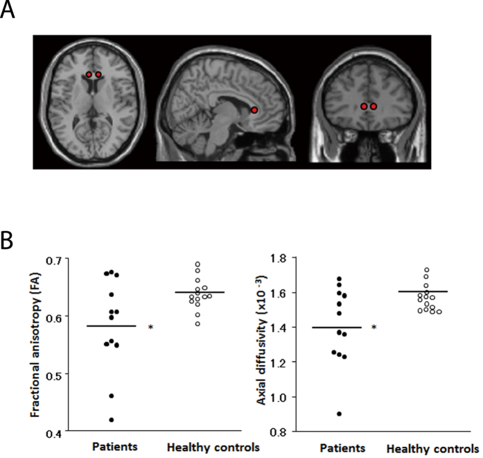
Figure 7. Lower fractional anisotropy (FA) values are observed in patients with MDD compared to healthy control subjects by DTI.
(A) Spherical voxels of interest (VOIs) placed on the anterior genu of the corpus callosum and (B) scatter plots of FA and axial diffusivity values in this region are shown for patients with MDD and controls. *Significantly lower FA and axial diffusivity values were observed in patients than in controls. Values were adjusted to the mean values of age and gender.

