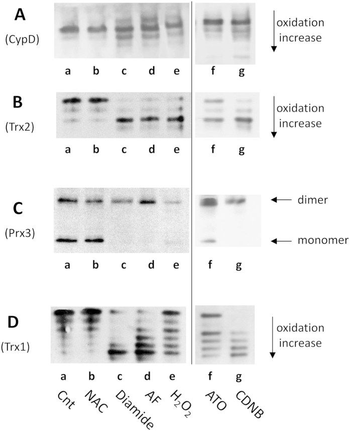
Figure 4. Redox Western blot of CypD, Trx1, Trx2 and Prx3 in CEM-R cells.
CEM-R cells (1 × 10⁶), treated for 18 h in various conditions, were derivatized with 10 mM IAM and then with 30 mM IAA for the determination of redox state of CypD, Trx1, Trx2 using urea-PAGE in non-reducing conditions (A–D). For the estimation of the redox state of Prx3, cell lysates were derivatized with 10 mM AIS and subjected to SDS-PAGE in non-reducing conditions (C). (a) control; (b) 2 mM NAC; (c) 2 mM diamide, (d) 3 μM AF; (e) 1 mM H2O2; (f) 15 μM ATO; (g) 20 μM CDNB.

