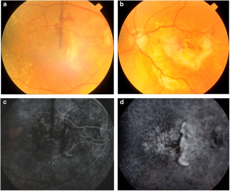
Figure 1.
Fundus photographs of patient 2 taken at the onset of right eye visual symptoms. Haemorrhage at the right macula (a), disciform scarring in the left eye (b) and bilateral drusen are shown. Early (c) and late (d) fluorescein angiography in the right eye revealed peripapillary choroidal neovascularisation.

