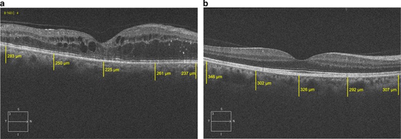
Figure 1.
OCT image of a DME patient (a) and a healthy control (b) on Cirrus. White lines show choroidal thickness measurements retrieved perpendicularly from the outer edge of the hyper-reflective retinal pigment epithelium. It was measured at five points; the subfoveal area, and the temporal and nasal points at a radius of 1 and 3 mm.

