Fig. 1.

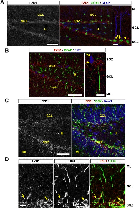
FZD1 is expressed in NSCs, amplifying progenitors and immature neurons of the adult mouse hippocampus. a Representative immunodetection of FZD1, SOX2 and GFAP in the dentate gyrus of 2-month-old mouse. Scale bar: 50 μm. Right, higher magnification of the image. Arrows indicate SOX2-positive cells with a single GFAP-positive projection. Arrowhead indicates a cell only positive for SOX2. Scale bar: 5 μm. b Immunodetection of FZD1, GFAP and the mitotic marker Ki67, scale bar: 50 μm. Right, higher magnification of the image. Arrows indicate cells positive for FZD1 and Ki67 staining. Scale bar: 10 μm. c Representative immunodetection of FZD1, DCX and NeuN in the dentate gyrus of 2-month-old mouse. Scale bar: 50 μm. d Higher magnification of the image shown in c. For simplification, only the double staining FZD1/DCX is shown. Arrows indicate FZD1-staining in the cell body of DCX-positive immature neurons. Scale bar: 10 μm. ML: molecular layer, GCL: granule cell layer, SGZ: subgranular zone, H: hilus
