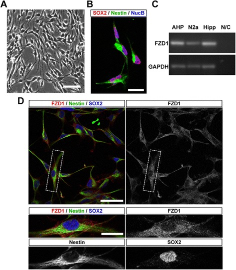
Fig. 2.

FZD1 is expressed in AHPs isolated from adult mouse hippocampus. a Monolayer of AHPs isolated from 6-week-old mouse hippocampus and cultured under proliferation conditions. Scale bar: 50 μm. b Immunoflurescence staining of SOX2 and nestin in AHPs. Nuclei were stained with NucBlue (NucB). Scale bar: 10 μm. c RT-PCR analysis of the expression of FZD1 and GAPDH in cultured AHPs (lane 1), N2a cells (lane 2), mouse hippocampus used as positive control (lane 3) and water used as PCR negative control (N/C, lane 4). d Immunoflurescence staining of FZD1, nestin and SOX2 in AHPs. Scale bar: 50 μm. Bottom panels, higher magnification of the cells indicated in top panels. Scale bar: 20 μm
