Fig. 5.

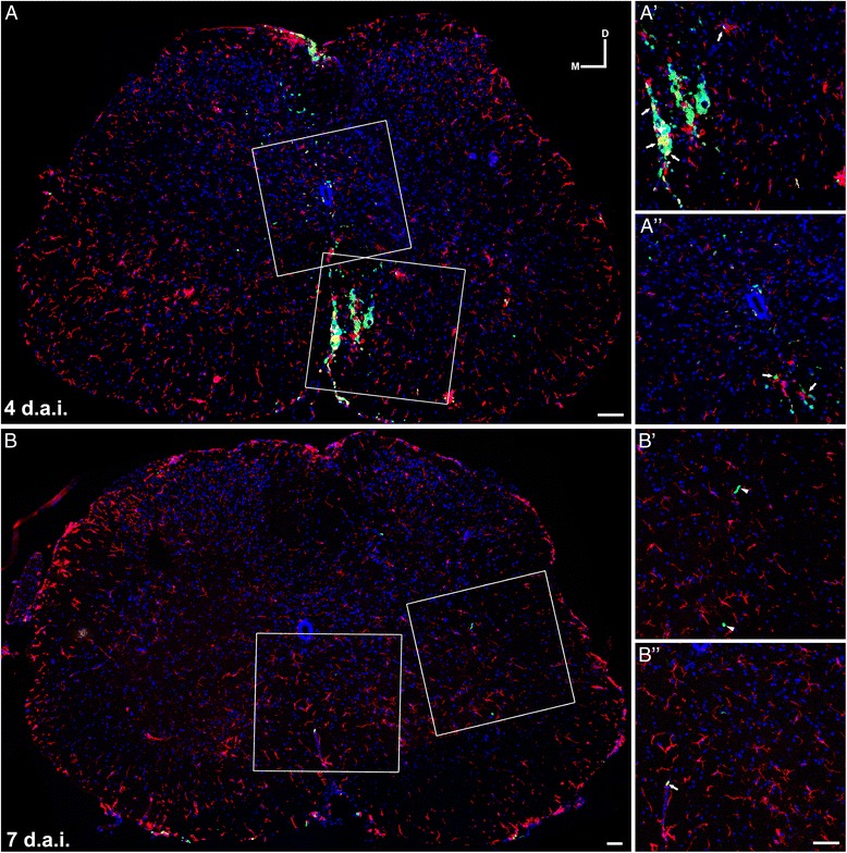
Fate tracing of BMMC-injected cells. BMMC expressing eGFP under the actin promoter were transplanted into the spinal cord of SOD1G93A mice in week 9 a, b. a Photomontage of confocal images showing a transverse section of lumbar spinal cord 4 days after BMMC transplant. Observe the wide distribution of BMMC-eGFP+ (green) on the dorsal–ventral axis. White boxes in a show in higher magnification (a′, a″) the association of BMMC with microglia stained with Iba1 (red). Arrows (a′, a″) indicate transplanted cells associated with microglia. b Photomontage of confocal images showing a transverse section of lumbar spinal cord 7 days after BMMC transplant. There was a sharp decrease in the number of BMMC-eGFP+ in the spinal cord. Some of the few remaining cells were not clearly associated with microglia (arrowheads in b′). Arrows (b″) indicate one of the few transplanted cells associated with microglia. b′, b″ show a higher magnification of white boxes in b. Scale bar: 50 μm. d.a.i. days after injection, D dorsal, M medial
