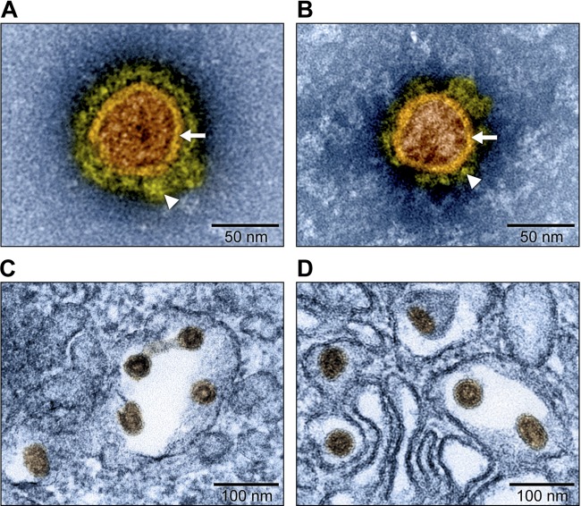
FIG 1 .
Electron micrographs of KRCV-1 and SHFV particles (artificially colored). Grivet kidney MARC-145 and MA-104 cells were infected with Kibale red colobus virus 1 (KRCV-1) and simian hemorrhagic fever virus (SHFV), respectively. (A) Electron micrograph of a negatively stained KRCV-1 particle from direct-pelleted supernatant. (B) Electron micrograph of a negatively stained SHFV particle from direct-pelleted supernatant. Samples were stained with 1.0% phosphotungstic acid. Note viral envelope (arrows) and envelope fringe proteins (arrowheads). (C) Electron micrograph of KRCV-1 particles in infected cells. (D) Electron micrograph of SHFV particles in infected cells.

