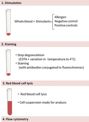
Fig. 1.

Diagram of the laboratory procedure for the basophil activation test. Following stimulation of blood cells with allergen or controls, blood cells are stained with antibodies coupled to a fluorochrome, which allow the identification of cells and the measurement of the expression of activation markers using a flow cytometer
