
Fig. 5.
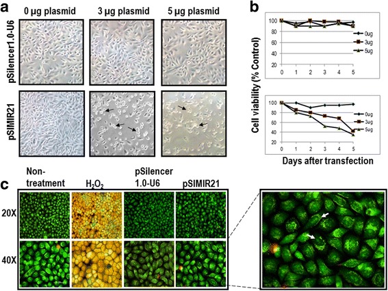
Analysis of tumor cell viability for silencing of miR-21 by siRNAs. Panel a SiHa cells were analyzed by white light microscopy (20X) 48 h after transfection with pSIMIR21 plasmid. The black arrows indicate the dead cells. Panel b Cellular viability was measured using MTS assay kit. Panel c SiHa cells attached on a slide were harvested at 48 h after transfection and stained with 5 μg/ml of acridine orange and propidium iodide dyes. Apoptosis control was induced by 5 μM of H2O2 added to cell culture during 2 h. A Nikon Elipse 400 epifluorescence microscope was used and samples were analyzed by FITC/TRITC using the 20X and 40X Fluor objectives. The white arrows indicate nucleus fragmented