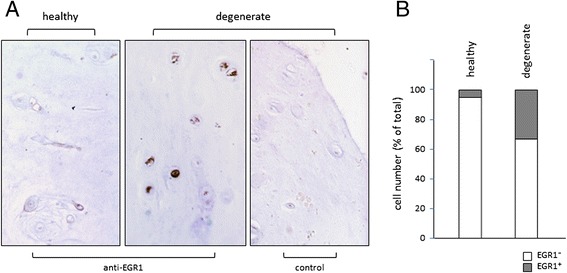
Fig. 5.

EGR1 expression is increased in degenerate NP cells. a Immunodetection of EGR1-positive cells in healthy control NP tissue (L1/L2 IVD of a 63 years old donor; left panel) and in degenerate NP tissue (L4/L5 IVD of the same donor middle panel); right panel: staining control on degenerate NP section: primary antiserum detecting EGR1 was left out. b Quantification of EGR+ cell numbers in healthy or degenerate NP tissue. Cell numbers are depicted as percentage of total cell number scored (total cell counts: 121 for healthy tissue and 218 for degenerate tissue)
