
Fig. 3.
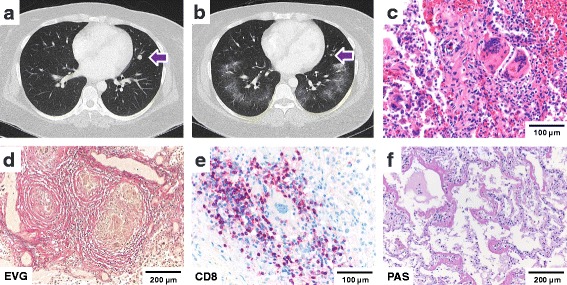
Lung damage patterns. a CT Thorax: Pulmonary metastasis before therapy with Ipilimumab (02/2015) b CT Thorax: pulmonary metastasis regression, ground glass opacities after Ipilimumab (04/2015) c sarcoid-like reaction d elastica stain showing epithelioid granulomas surrounded by fibrotic rings e CD8-positive T-cell infiltrates surrounding giant cell granulomas as detected by immunohistochemistry f diffuse alveolar damage; scale bars as indicated