
Fig. 1.
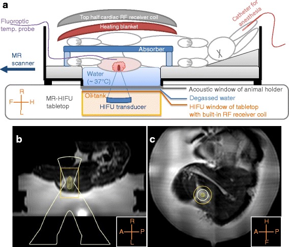
Experimental setup. A schematic overview of the experimental setup is shown in (a). The animal holder was placed with its acoustic window above the HIFU window; degassed water was used for acoustic coupling. The shaved tumor-bearing leg was positioned above the acoustic window, and a fluoroptic temperature probe was inserted in the tumor-bearing leg, in the far-field of the HIFU beam. The tank was filled with warm water (~37 °C) up to the tumor-bearing leg, and an absorber was placed between the legs. On top of the rabbit, a heating blanket and a flat 16-channel array coil was placed. In (b, c), examples of the treatment planning are shown on reconstructed sagittal and coronal images of the T2-weighted 3D turbo spin-echo acquisition