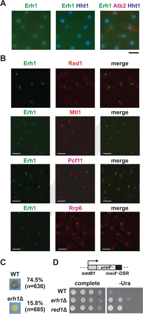
Figure 1. Characterization of Erh1.
(A) Erh1 forms nuclear foci. Representative images of vegetative fission yeast cells expressing Erh1-GFP (Erh1), Hht1 (histone H3 h3.1)-CFP and mCherry-Atb2 (tubulin alpha2; used to confirm mitotic interphase) are shown. Bar, 5 μm. (B) Erh1 colocalizes with Red1, Mtl1, Pcf11 and Rrp6. Deconvolved images of vegetative fission yeast cells expressing Erh1-GFP (Erh1) with either Red1-tdTomato (Red1), Mtl1-tdTomato (Mtl1), Pcf11-mCherry (Pcf11) or Rrp6-mCherry (Rrp6) are shown. Bars, 5 μm. (C) Cells from a parental WT or erh1Δ homothallic culture were spotted onto minimal (PMG) plates and incubated at 26°C for 3 days. The presence or absence of asci was determined by iodine staining, and the mating efficiencies are noted; “n” indicates the number of cells counted in each strain. (D) Erh1 is required for DSR-dependent mRNA decay. The ura4+-DSR construct is shown (top). WT, erh1Δ, and red1Δ cells carrying the ura4+-DSR were spotted onto complete or uracil-lacking (-ura) minimal plates in the absence of thiamine and then were incubated at 30°C for 3 days (bottom). See also Figure S1.

