Figure 5.
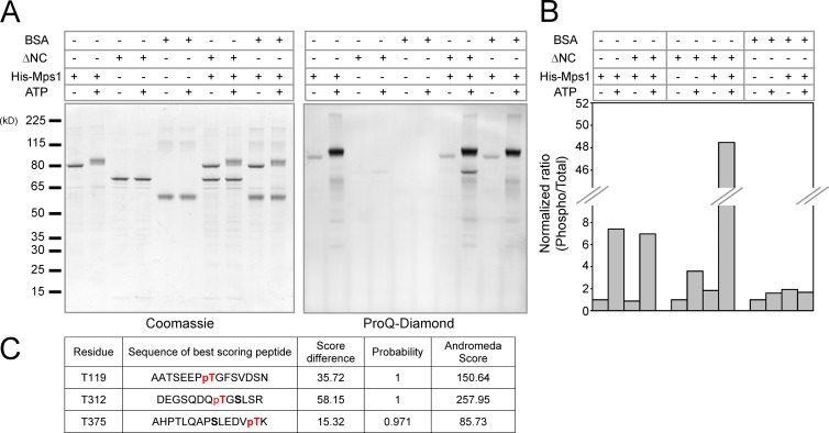
Mps1 phosphorylates ARHGEF17 in vitro. (A) In vitro kinase assay of Mps1 and ARHGEF17. Recombinant His-tag fused Mps1 (kinase) and untagged ARHGEF17-ΔNC or BSA (substrate) were incubated in the presence or absence of ATP. Total protein was visualized with Coomassie brilliant blue (CBB; left), and phosphorylated protein was visualized with Pro-Q Diamond (right). (B) Comparison of normalized mean intensity ratio between phosphorylated protein and total protein in each condition. Quantification was performed from single experiment. (C) Potential phosphorylation sites of ARHGEF17 by Mps1 identified by LC-MS/MS. Andromeda score, probability, and delta score are indicated for each site (see Materials and methods).

