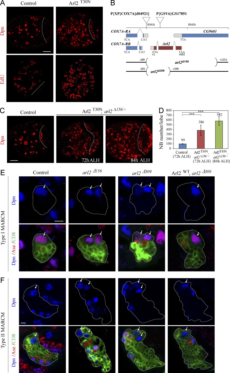
Figure 1.
Loss of arl2 results in ectopic NBs in Drosophila larval brains. (A) Control and Arl2T30N larval brains labeled for Dpn and EdU. (B) A schematic diagram of deleted regions in arl2Δ156 and arl2Δ309, together with the location of COX7A and P[GSV6]GS17851. (C) Larval brain from a control at 72 h ALH, Arl2T30N, arl2Δ156/+ (overexpressed Arl2T30N in arl2Δ156/+) at 72 h ALH and 84h ALH labeled for Dpn. Central brain is to the left of the white dotted line in A–C. (D) Quantification of central brain NB number per brain hemisphere (with SD) in C: control (72h ALH), 98.6 ± 5.0 (n = 23); Arl2T30N, arl2Δ156/+ (72h ALH), 385.6 ± 107.6 (n = 25); Arl2T30N, arl2Δ156/+ (84h ALH), 581.5 ± 93.9 (n = 25). ***, P < 0.001. (E and F) Type I (E) and II (F) NB MARCM clones in control (FRT82B), arl2Δ156, arl2Δ309, and Arl2WT overexpression in arl2Δ309 labeled for Dpn, Ase, and CD8. Cells in the clones are labeled by CD8-GFP, and outline of the NB lineages is indicated by white dotted lines. Arrows indicate NBs. Bars: (A and C) 20 µm; (E and F) 5 µm.

