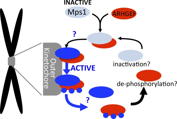
Figure 1.
Proposed timer model for Mps1 retention at kinetochores. ARHGEF17 (red) binds inactive Mps1 (light blue) in the cytoplasm, and the complex binds to the outer kinetochore (gray) where Mps1 becomes activated (blue). Mps1 phosphorylates ARHGEF17 (blue circles), causing dissociation of both from kinetochores. It is still unknown where and how Mps1 is activated upon ARHGEF17 binding or where ARHGEF17 dissociates from Mps1 (blue question marks), and if and how Mps1 is inactivated or ARHGEF17 is dephosphorylated upon returning to the cytoplasm.

