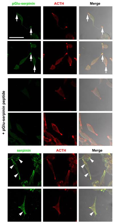
Fig. 2.
Intracellular distribution of pGlu-serpinin in AtT-20 cells. The images show that immuno-reactive pGlu-serpinin (Alexa 488, green) was detected mainly in the tips of AtT-20 cells (arrows), and co-localized (merged, yellow) with a secretory granule marker, ACTH (Cy3, red) (upper panels), whereas no immunosignal was detected in the absorption control group (+pGlu-serpinin peptide, middle panels), which was incubated with anti-pGlu-serpinin antibody in the presence of synthetic pGlu-serpinin peptide. In contrast (lower panels), immunore-active serpinin/CgA detected by anti-serpinin antibody (Alexa 488, green) was distributed not only in the tips, but also in the processes and soma (arrow heads) of AtT-20 cells. Scale bar=50 μm

