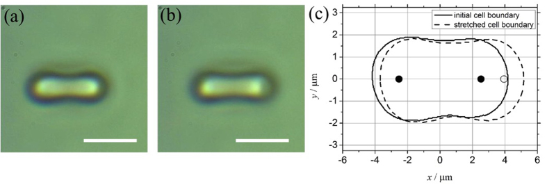
Figure 2. Side on images of RBC under dual beam optical tweezer set up.
(2a) Unstretched RBC: Side on view of a unstretched RBC (scale bar: 5 um). (2b) Stretched RBC: Side on view of a Stretched RBC (scale bar: 5 um). (2c) RBC boundaries demarcated – continuous edges representing unstretched RBC, broken edges representing stretched RBC, black dots indicates laser spots at the start and white dots indicate the laser spot position at the time of maximum stretch.

