
Figure 2. Immunohistochemical analysis of tissue factor expression at the decidual–placental interface.
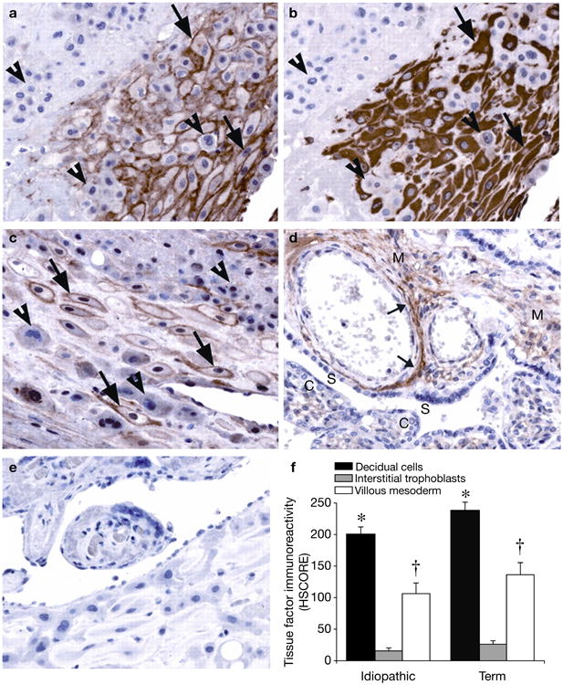
Serial sections of decidual basalis specimens immunostained for tissue factor (TF) and vimentin, as shown in an idiopathic preterm specimen: (a) TF and (b) vimentin. Decidual cells (arrows), identified by positive vimentin staining, exhibit strong perimembranous TF staining. TF staining is absent in the interstitial trophoblast (arrowheads). Similar results are seen in term specimens (c). Placental villi show moderate TF staining in the mesoderm (M), in which the staining is primarily localised to perivascular adventitia (small arrows), as shown in a preterm specimen (d). The syncytiotropoblast (S) and cytotrophoblast (C) show no TF staining. Similar results are seen in term specimens (not shown). Negative control immunostaining using a nonspecific isotype-matched antibody revealed no positive signals (e). TF staining intensity (f) [bars indicate histological score (HSCORE) mean ± s.e.m.] is highest in decidual cells (*P<0.05, versus interstitial trophoblast and the villous mesoderm of the same specimen group), with moderate staining in the villous mesoderm (†P<0.05, versus interstitial trophoblast of the same specimen group). There are no significant differences in HSCORE values between specimen groups. Published with permission from Ref. 46 (© 2009, The Endocrine Society).