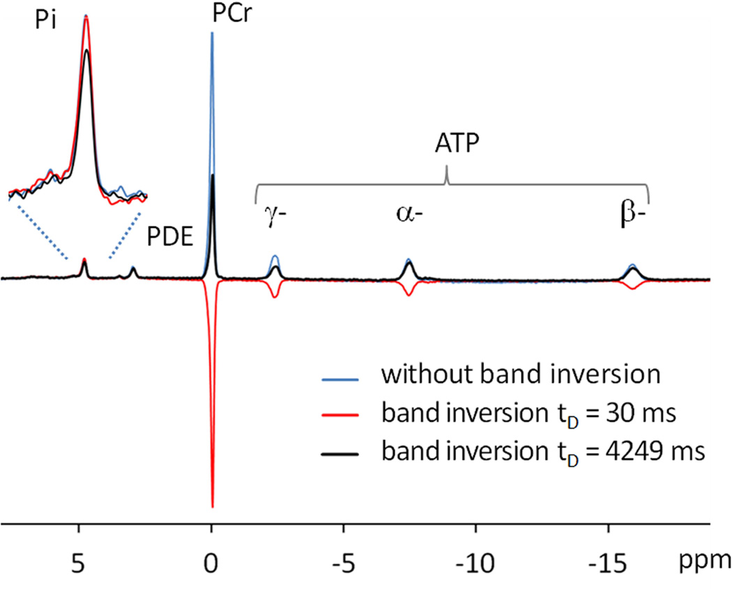
FIG. 3.
Representative 31P MR spectra acquired from resting human calf muscle at 7 T using a single pulse without inversion (blue trace) and using EBIT sequence with delay time tD at 30 ms (red trace) and at 4249 ms (black trace). All spectra were collected with TR = 30 sec and NA = 4 with identical y-scale.

