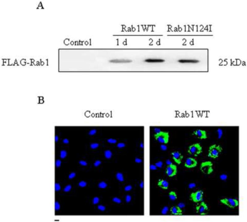
Fig. 3.
Lentivirus-mediated expression of Rab1 in RPMVECs. (A) Western blot analysis of expression of wild-type Rab1 (WT) and dominant-negative mutant Rab1N124I driven by lentivirus. RPMVECs were infected with empty lentivirus vector (Control) or recombinant FLAG-Rab1WT lentivirus for 1 and 2 days or with Rab1N124I for 2 days at an m.o.i. of 20. 40 μg of whole RPMVECs was separated by 12% SDS-PAGE, and FLAG-Rab1 expression was detected by immunoblotting with anti-FLAG antibody. The immunoblot is representative of results obtained in two different experiments. (B) localization of Rab1WT and estimation of infection efficiency. RPMVECs were cultured on coverslips and infected with control or GFP-Rab1WT lentivirus. Localization of GFP-Rab1WT in infected RPMVECs was revealed by fluorescence microscopy as described in the Materials and methods section. Similar results were obtained in three separate experiments.

