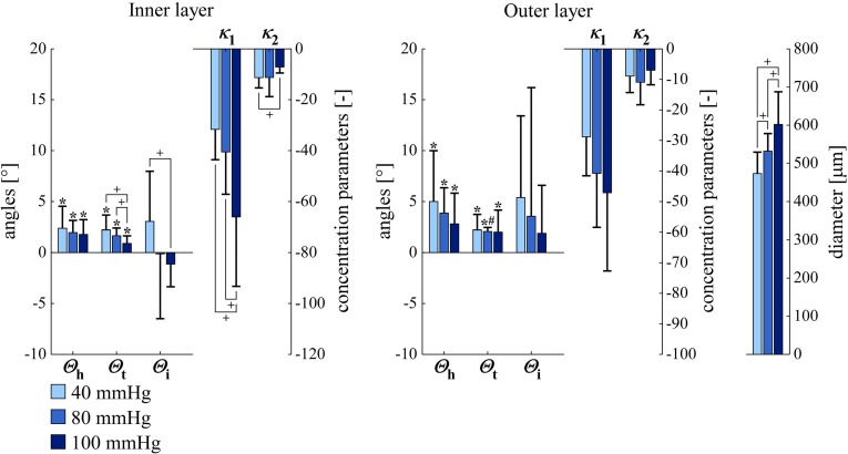
Fig. 8.
SMC orientations and arterial diameters in left carotid arteries at luminal pressures of 40, 80, and ( for , , and ; for , , and diameter). versus angle zero (Rayleigh test). inner versus outer layer (Rayleigh or paired Student’s t test). for parameter difference between pressures (Rayleigh or paired Student’s t test). , , , , and are defined in Fig. 6. SD standard deviation, SMC smooth muscle cell

