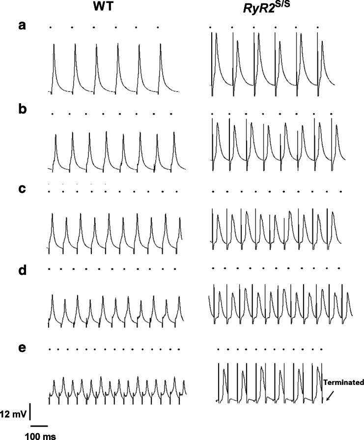
Fig. 3.
Typical MAP recordings obtained from the left ventricular epicardium of WT and RyR2 S/S during dynamic pacing. Traces from WT (left) and RyR2 S/S at progressively decreasing BCLs: 124 (a), 99 (b), 84 (c), 74 (d) and 54 ms (e). If a heart entered 2:1 block, the protocol was terminated (E). Traces are displayed along a common horizontal timescale. The vertical scale was normalized to a standard AP deflection at a BCL of 134 ms. Small black circles above each trace indicate the timing of stimuli

