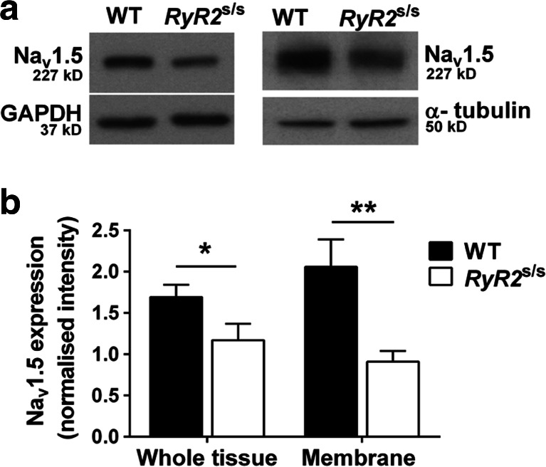
Fig. 7.
Western blots of Nav1.5 expression in whole tissue and membrane fraction samples from WT and RyR2 S/S ventricles. Ventricular Nav1.5 expression was decreased in RyR2 S/S compared to WT, both in the whole tissue (1.17 ± 0.20; n = 6, vs 1.69 ± 0.15 n = 7, respectively, P = 0.048) and in the membrane fraction (0.91 ± 0.13; n = 4, vs 2.06 ± 0.33; n = 4, respectively, P = 0.006). This suggested a greater proportional reduction in membrane relative to total Nav1.5 expression in RyR2 S/S. Symbols denote significant differences between genotypes *P < 0.05, **P < 0.01

