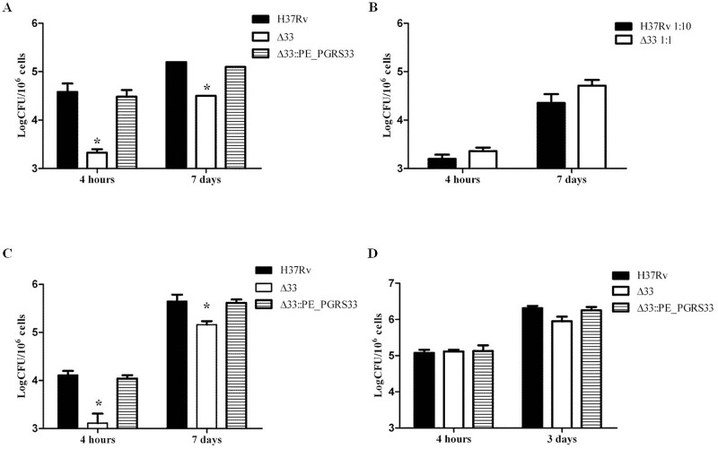
Fig 1. MtbΔ33 is impaired in its ability to enter macrophages.
Murine J774 macrophages (A and B), human monocyte-derived macrophages THP-1 (C) and human type II pneumocytes A549 (D) were infected with Mtb H37Rv, MtbΔ33 and MtbΔ33::PE_PGRS33 strains at a MOI 1:10 (A-C) or 5:1 (D) and following incubation cells were washed and at the different time points intracellular bacteria were determined by CFU counting. In the experiment shown in panel B, the Mtb H37Rv was infected at MOI 1:10, while the MtbΔ33 at MOI 1:1. Results of one representative experiments from at least three assays are shown (*p<0.,01). CFUs were expressed as mean ± SD and were analysed by two-way ANOVA followed by Bonferroni posttest.

