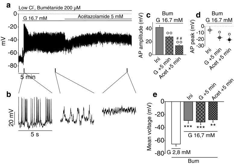
Fig. 8.
Effect of glucose on membrane potential oscillations in single rat β-cells exposed to low Cl−/HCO3 −-free medium. Zero-current nystatin-perforated patch-clamp voltage recordings. Sampling rate, 18 kHz; 2-kHz filter setting. Dotted lines represent zero-voltage level. β-Cells were continuously exposed to 200 μM bumetanide (Bum), a NKCC1/2 inhibitor, and stimulated with 16.7 mM glucose (G 16.7 mM) in Hepes-buffered NaCl solution without bicarbonate containing 20 mM Cl−. After 10-min glucose stimulation, 5 mM acetazolamide (Acet), a carbonic anhydrase inhibitor, was added to further reduce intracellular HCO3 −. a Representative recording. b Extended timescale of recording a. c AP amplitude at various times, i.e., at the beginning of the spiking activity before rundown (Ini), after 5 min in presence of glucose stimulation (G + 5 min), and after 5 min in presence of acetazolamide (Acet + 5 min). d AP peak voltage at similar times. e Mean voltage at similar times. For all results, n = 6 experiments performed on one preparation of rat islets. Repeated measures ANOVA test on c, P = 2.50 × 10⁻⁵; d, P = 1.29 × 10⁻⁴; and e, P = 7.42 × 10⁻¹⁰. **P < 0.01, ***P < 0.001 vs. 2.8-mM glucose condition in presence of bumetanide; °P < 0.05, °°P < 0.01 vs. Ini; ##P < 0.01 vs. G + 5 min (Sidak tests)

