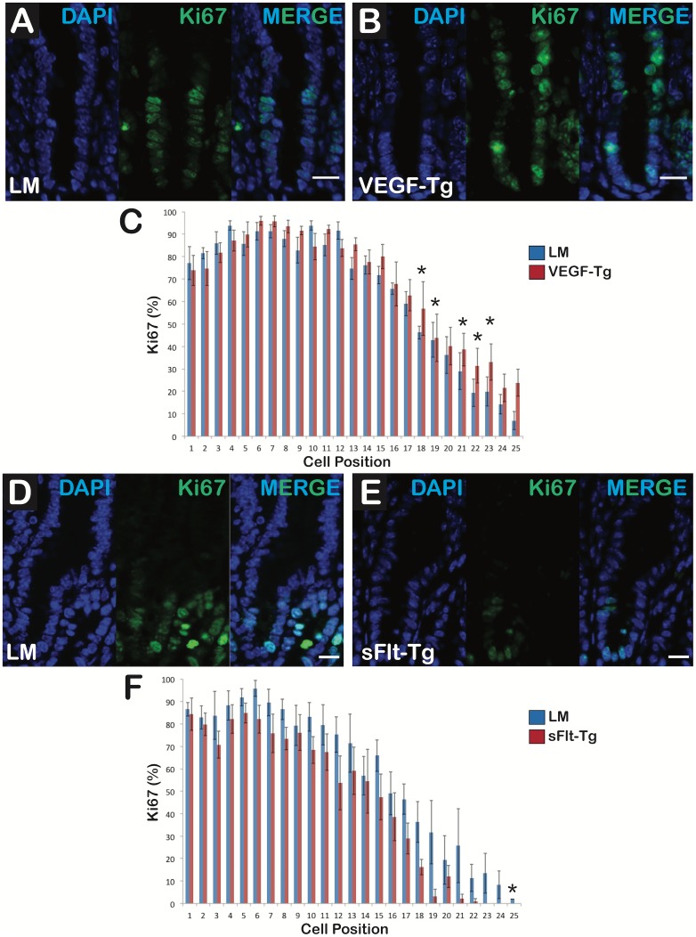
Fig 6. VEGF augmentation increased epithelial cell proliferation in the transit-amplifying zone whereas reduced VEGF bioavailabity restricted proliferation to the crypt bases.
(A) Littermate crypts had typical proliferation in the crypt and transit-amplifying zone of the duodenum. (B) VEGF mutant crypts displayed increased proliferation at higher positions in the transit-amplifying zone compared to littermates. First panel shows nuclei with DAPI staining (Blue), second reveals Ki-67-positive cells (Green) and third panel is the composite (MERGE); N = 6 mice per group; Scale bars = 20 μm (C) VEGF mutants had more Ki-67-positive cells at positions 16 through 25 than the littermates. (D) Littermate crypts had typical proliferation in the crypt and transit-amplifying zone. (E) sFlt-1 mutant crypts displayed decreased proliferation in the transit-amplifying zone compared to littermates. First panel shows DAPI staining (Blue), second reveals Ki-67-positive cells (Green) and third is the composite (MERGE); N = 6 mice per group; Scale bars = 10 μm (F) sFlt-1 mutants had fewer Ki-67-positive cells at positions 10 through 25 than littermates. N = 6 mice per group; *p<0.05; Error bars = SEM.

