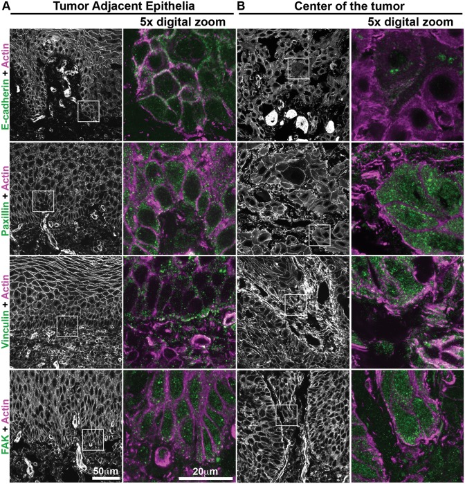
Fig 5. Human oral squamous cell carcinoma biopsies show differential distribution of adhesion proteins between center of the tumor cells and tumor-adjacent epithelia.
Regions of biopsies corresponding to the epithelia adjacent to the tumor (A) and from the center of the tumor (B) were submitted to immunostaining for E-cadherin, paxillin, vinculin or FAK (green) and actin staining (magenta). Inserts demonstrated in actin staining, were digitally magnified (5x) to show intracellular localization. Representative images from different patients (n = 10), scale bar = 50μm or 20μm.

