
Figure 4. Upregulation of miR-181c promotes pancreatic cancer cell chemoresistance in vitro.
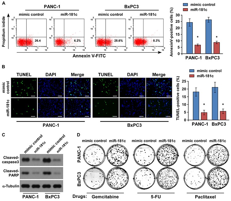
A. Annexin V-FITC/PI staining of indicated cells treated with gemcitabine (5 μM) for 24 h. B. Representative micrographs (left) and quantification (right) of TUNEL-positive cells following 36-h gemcitabine (5 μM) treatment. Scale bars: 50 μm. C. Western blotting of cleaved caspase3 and PARP expression. α-Tubulin was used as the loading control. D. Representative micrographs of crystal violet–stained PANC-1 and BxPC3 pancreatic cancer cell colonies in the presence of gemcitabine (5 μM), 5-FU (5 μM), or paclitaxel (10 μM). Error bars represent the mean ± s.d. of three independent experiments. *P < 0.05.