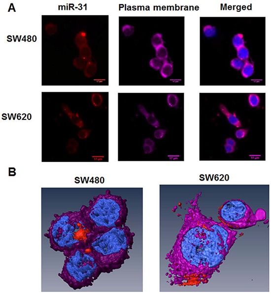
Figure 1. Distribution of miR-31 molecules in SW480 and SW620 CRC cells by conventional microscopy, including 3D-reconstruction of confocal images.

A. Conventional microscopy images of SW480 and SW620 cells. The human CRC SW480 (low metastatic potential) and SW620 (highly metastatic) cell lines were transfected with 10 nM of miR-31 probe-Alexa568 (red color) for 24 h. Then, the plasma membranes of cells were stained with Cell Mask Deep Red (purple color). Cells were fixed by 4% PFA and nuclei were stained with DAPI (blue color). B. 3D reconstruction of selected cells from (A) above.
