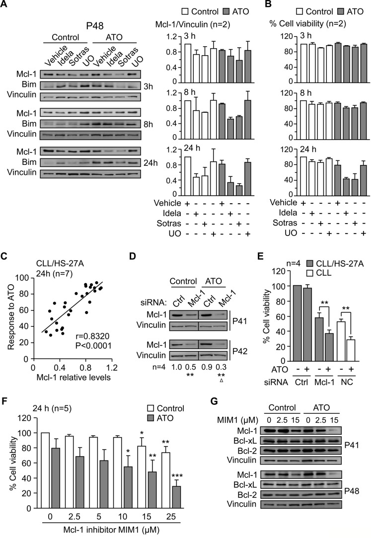
Figure 7. Mcl-1 plays a central role in the mechanism of CLL cell resistance to ATO induced by stroma.
(A) 8–10 × 106 CLL cells were treated with kinase inhibitors as in Figure 6A and cultured on HS-27A cells, with or without 2 μM ATO, for the indicated times. Western blotting analyses are shown for a representative patient and Mcl-1 values quantitated for the two samples analyzed (P41, P48). (B) Average cell viability of the same samples used in (A). (C) CLL cells (P10, P32, P35, P40, P41, P42, P48) were treated for 1 h with vehicle, 5 μM UO, idelalisib or sotrastaurin (2.5 and 25 μM each), and cultured on HS-27A cells with 2 μM ATO. Cell viability and Mcl-1 expression were determined after 24 h. Pearson's correlation (r) and P values are shown. (D, E) 24 × 106 CLL cells (P32, P40, P41, P42) were nucleofected with the indicated siRNAs and cultured in suspension or with HS-27A cells. After 16 h at 37°C, 2 μM ATO was added and cells further incubated for 24 h. Nucleofected samples were analyzed by Western blotting (D) and cell viability determined by flow cytometry (E). NC (nucleofection control): CLL cells nucleofected with control siRNA and cultured in suspension. (F) CLL cells (P32, P41, P42, P43, P48) were treated for 1 h with or without the indicated concentrations of MIM1 and cultured with HS-27A cells in the absence or presence of 2 μM ATO. After 24 h, cell viability was determined by flow cytometry. (G) 8–10 × 106 CLL cells were treated with the indicated concentrations of MIM1 and cultured as in (F). Protein expression was analyzed by Western blotting. *or Δ P ≤ 0.05; **or ΔΔ P ≤ 0.01; ***or ΔΔΔ P ≤ 0.001.

