Fig. 1.
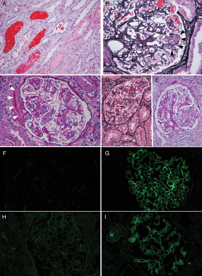
Pathology of allograft biopsies and native nephrectomy, Patient 1. (A) Biopsy Post-transplant Day 96 showing numerous RBC casts and tubular injury. (B) Biopsy Post-transplant Day 96, glomerulus with segmental endocapillary hypercellularity (black arrowheads). (C) Biopsy Post-transplant Day 342, glomerulus with segmental mesangial and endocapillary hypercellularity. White arrowheads highlight segmental fibrosis, consistent with fibrocellular crescent. There were also prominent RBC casts in tubules (not shown). (D) Native nephrectomy specimen showing glomerulus with endocapillary proliferation and reactive appearing extracapillary cells. (E) Native nephrectomy specimen with segmental mesangial proliferation. (F-I) Immunofluorescence microscopy. Each of the tested biopsies showed ultrathin linear glomerular capillary loop staining for IgG3 and lambda. IgG1 and kappa were negative. IgG2 and IgG4 were also negative (not shown). (F) IgG1 immunofluorescence, Day 342. (G) IgG3 immunofluorescence, Day 342. (H) Kappa light-chain immunofluorescence, Day 160. (I) Lambda light-chain immunofluorescence, Day 160. Hematoxylin & eosin (H&E) stain panel A; Jones silver stain panels B and D; Periodic acid Schiff (PAS) stain panel.

