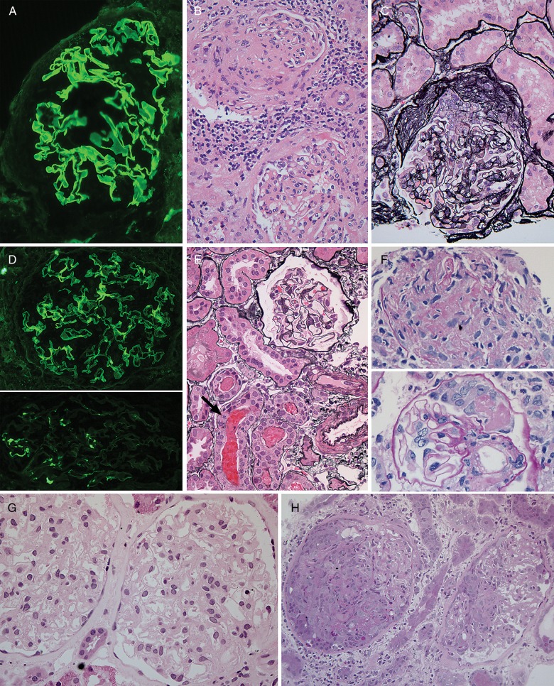
Fig. 2.
Histopathologic findings in atypical anti-GBM disease. (A-C) Patient 2. (A) IgG immunofluorescence staining shows strong, linear staining of GBMs. (B) H&E stain showing segmental glomerular scar (top), interstitial inflammation (middle) and glomerulus with mesangial expansion and tuft adhesions (bottom). (C) Jones silver stain shows fibrocellular crescent. (D-F) Patient 3. (D) IgG immunofluorescence staining shows strong, linear staining of GBMs in top panel. Bottom, IgA with granular mesangial staining. (E) The majority of glomeruli, as shown here with Jones silver stain, were histologically normal. RBC casts were seen, at bottom. Arrow denotes tubular epithelial cell with mitotic figure. (F) The only two abnormal glomerular foci, not present on initial levels, including fibrin-inflammatory focus (top panel), and small segmental crescent with fibrin (bottom panel), PAS stain. (G-H) Patient 5. (G) First biopsy with histologically normal glomeruli (H&E) despite strong linear IgG immunofluorescence staining (not shown). (H) Biopsy 3 months later with necrotizing and crescentic glomerulonephritis (PAS stain). Original magnifications: 200× B–C, H; 400×: A, D–G.

