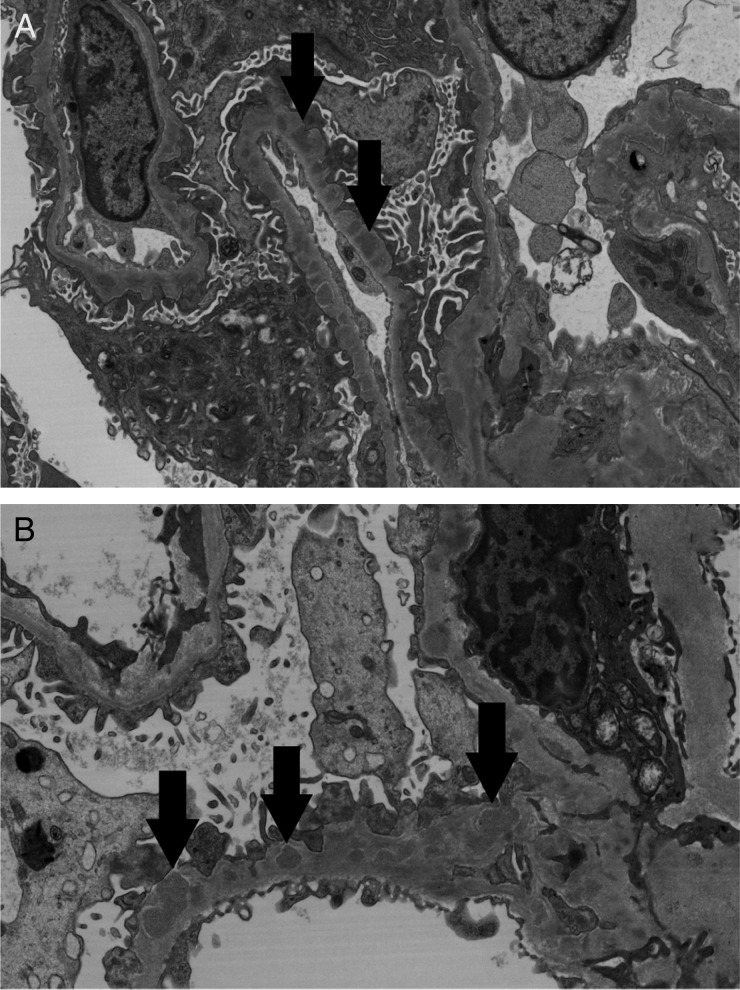
Fig. 2.
(A and B) Electron micrographs show numerous immune-type electron-dense deposits in peripheral capillary walls in intramembranous and subepithelial locations (arrows). There are ‘spikes’ of basement membrane matrix separating the deposits and incorporating them into the basement membranes [original magnification: (A) ×7960; (B) ×11900].

