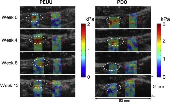
Figure 4. Elastography imaging of engineered scaffolds in vivo.
Shear modulus images of degradable poly(ester urethane)urea (PEUU) and polydioxanone (PDO) scaffolds implanted in rat abdominal wall. White and red circles indicate regions of the scaffold and native abdominal wall, respectively. Reprinted from Biomaterials, 35/27, Park D.W., Ye S-H, Jiang H.B., Dutta D., Nonaka K., Wagner W.R., Kim K., In vivo monitoring of structural and mechanical changes of tissue scaffolds by multi-modality imaging, 7851, 2014, with permission from Elsevier.

