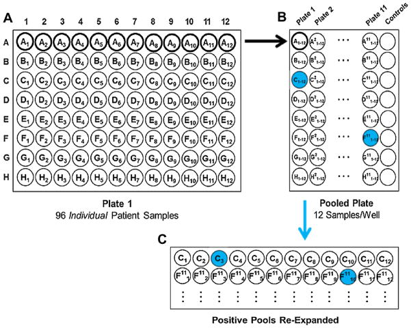
Fig. 1.
Specimen pooling and screening schematic. (a) Plate 1 is an 8 × 12 plate with individual DNA samples. Each row was combined into 1 well in the (b) pooled plate, which was screened for HHV-6 with qPCR (high positive pools are shaded). (c) Individual samples from high positive pools were screened for HHV-6 by qPCR, and inherited ciHHV-6 was confirmed in high positive individual samples (shaded) using ddPCR.

