
Figure 3. Example of cell type-specific expression of DREADDs.
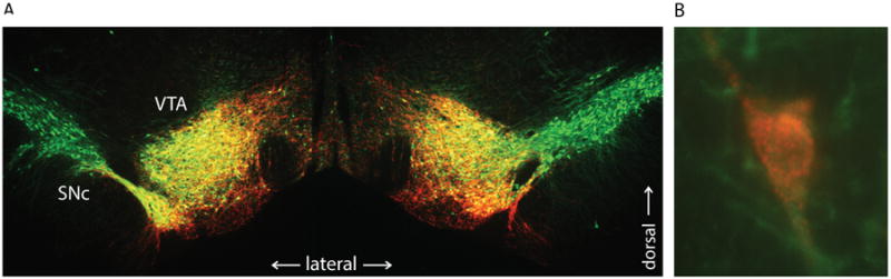
A, Tiled 5× coronal images showing bilateral expression of hM3Dq in the midbrain (AAV2-DIO vector in TH-Cre rat). mCherry (red; DS Red immunostain) shows DREADD expression superimposed on tyrosine hydroxylase-positive (TH+) neurons (green; TH immunostain) in the ventral midbrain. Overlap indicated in yellow, showing restricted DREADD expression in ventral tegmental area (VTA) and not in lateral substantia nigra pars compacta (SNc). B, 60× image of neuron co-expressing hM3Dq (red) and TH (green). Note membrane bound, punctate staining of mCherry-fused hM3Dq receptors.