
Figure 4. Example of axonal expression of DREADDs.
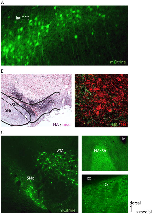
A, 10× coronal image of lateral orbitofrontal cortex (lat OFC) neurons that project to the striatum expressing the Gi DREADD (natural mCitrine fluorescence). CAV-Cre injections were made in a focal point of the striatum and Cre-dependent hM4Di delivered to the lat OFC. B, Left, 5× coronal image example of axonal expression (HA-tag immunostain, dark purple) of hM4Di in Nissl-counterstained ventral tegmental area (VTA) and substantia nigra pars compacta (SNc), following lentiviral syn-hM4Di injection in the rostral ventral pallidum (example pallidum injection in Fig. 1). Right, 60× confocal image showing comingling of hM4Di-expressing fibers (green; HA-tag immunostain) and tyrosine hydroxylase-positive (TH+) neurons (red; TH immunostain). C, Left, coronal image of midbrain VTA and SNc expressing hM4Di in TH+ neurons in the TH-Cre rat (natural mCitrine fluorescence). Right, labeled fibers in the nucleus accumbens (NAc; predominantly shell region; top) and fibers in the dorsal striatum (DS) arising from dopaminergic neurons (bottom) in the same brain. cc, corpus callosum. lv, lateral ventricle.