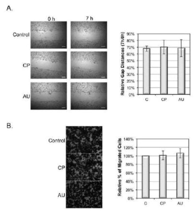
Fig. 5.
Effect of sialidase on migration and invasion of MDA-MB-231 cells. (A) MDA-MB-231 cells were grown on a fibronectin-coated plate to reach confluence and were treated with 0.1U/ml C. perfringens (CP), and A. ureafaciens (AU) sialidase in DPBS for 30 min at 37°C. The wound was then made using a 200 μl pipette tip. Cells were incubated in FBS free medium for 7 h at 37°C. Photographs were taken at 0 h and 7 h. Gap distances were shown as the ratio of 7 h to 0 h. Duplicate samples were prepared in each experiment. Data shown are the means ± SD, n=4. *: p<0.05 versus control group. Scale bar, 250 μm. (B) MDA-MB-231 cells were harvested and treated with sialidases as described in (A). 1×10⁵ MDA-MB-231 cells were seeded into inner chamber of transwell inserts coated with fibronectin (FN). After 24 h incubation at 37°C, cells migrated to the lower surface were fixed, stained and then counted under a microscope from 4 different fields. Duplicate samples were prepared in each experiment. Data shown are the means ± SD, n=3. *: p<0.05 versus control group. Scale bar, 50 μm.

