Figure 5.
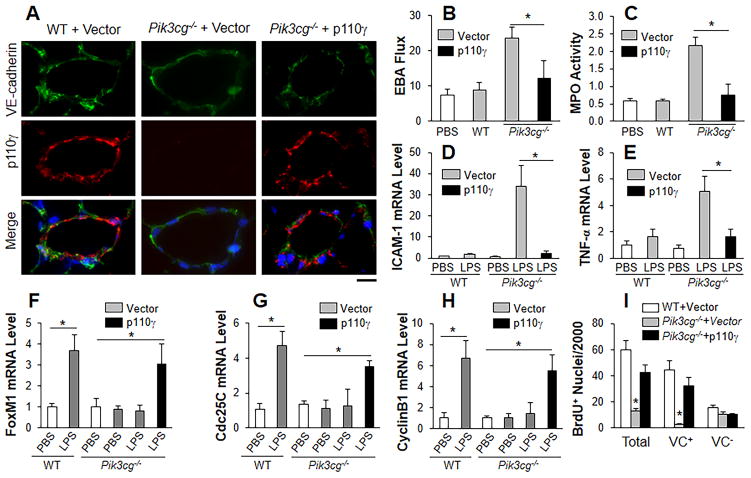
Restored expression of p110γ in lung endothelial cells of Pik3cg−/− mice induces FoxM1 expression and normalizes endothelial regeneration. (A) Representative micrographs showing endothelial expression of p110γ in pulmonary vascular ECs of Pik3cg−/− mice at 30h following liposome-mediated transduction of p110γ plasmid DNA (driven by human CDH5 promoter). Scale bar, 30μm. (B) Endothelial expression of p110γ normalized the defective vascular repair phenotype of Pik3cg−/− mouse lungs. At 30h post-plasmid DNA transduction, mice were challenged with LPS and lungs were collected for assessing pulmonary transvascular EBA flux at 60h post-LPS. n = 5. *, P < 0.01 (t test). (C) Lung tissue MPO activity indicating resolved lung inflammation in Pik3cg−/− mice transduced with p110γ plasmid DNA at 60h post-LPS. *, P < 0.01(t test). (D, E) QRT-PCR analysis of expression of pro-inflammatory molecules in mouse lungs. *, P < 0.01 (t test). (F–H) QRT-PCR analysis of FoxM1 expression and its downstream target genes in Pik3cg−/− lungs transduced with p110γ plasmid DNA. *, P < 0.01 (t test). (I) Endothelial expression of p110γ normalized lung EC proliferation in Pik3cg−/− mice at 60h post-LPS. *, P < 0.001 (ANOVA). VC, vWF/CD31.

