Figure 7.

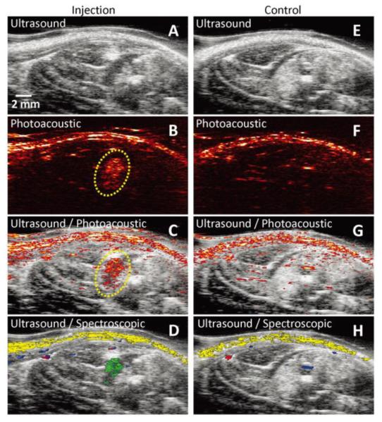
In vivo tracking of gold nanotracers (Au NT) labeled mesenchymal stem cells (MSCs) intramuscularly injected in the lower limb of the Lewis rat, using combined ultrasound and photoacoustic (US/PA) imaging. The ultrasound image shows the structural information of the lower limb, but the location of MSCs cannot be identified. However, the photoacoustic and US/PA images clearly show the location of the nanotracer signal from MSCs in the gel outlined in yellow (B). (A–D) In vivo ultrasound, photoacoustic, US/PA, and US/spectroscopic images of the lateral gastrocnemius (LGAS) in which PEGylated fibrin gel containing Au NT loaded MSCs (1×10⁵ cells/mL) was injected. The gel location is outlined with yellow dotted circle. Injection depth was about 5 mm under the skin. (E–H) Control at the region of the LGAS of the other hind limb without any injection. Spectral (650–920 nm) analysis of photoacoustic signal was able to differentiate skin (shown in yellow), oxygenated (red) and deoxygenated (blue) blood, and Au NT loaded MSCs (green). The images measure 23 mm laterally and 12.5 mm axially. Adapted from Fig. 6. 50
