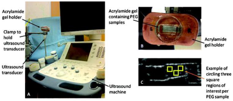
Figure 1.
Experimental setup. (A) Ultrasound imaging setup showing the custom made PEG hydrogel holder fixed above the transducer. (B) Custom made imaging platform consisting of a water-filled acrylamide mold containing PEG hydrogel samples. (C) Sample output image: for each set of samples, images were gathered for three different sagittal cross sections, and three regions of interest were manually chosen for each gel in the image.

