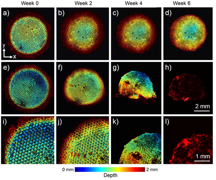
Figure 6.

(a–h) OR-PAM coronal projection images showing the degradation of a PLGA inverse opal scaffold immersed in (a–d) plain PBS and (e–h) PBS containing 0.025 wt.% lipase at 37 °C for a period up to 6 weeks. (i–l) Magnified views showing the top-left corner of the images in (e– h), respectively. While the scaffold in PBS did not undergo obvious structural alterations for up to Week 6, the scaffold showed remarkable changes over time in the presence of lipase. The images are color-coded by depth of maximum. Reprinted with permission from Ref.77, copyright 2014 Wiley-VCH.
