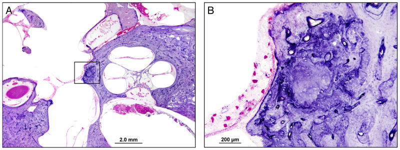
Figure 1.
Temporal Bone Case 1: A) Low power image through the cochlea, vestibule and oval window. No otosclerosis is seen. There is evidence of a stapedectomy with complete removal of the stapes footplate. B) High power image of the boxed area in A, which includes the otic capsule at the anterior margin of the oval window. There is basophilic staining anterior to the oval window with a prominent cartilaginous rest in the area of the fissula ante fenestram, but there is no otosclerosis.

