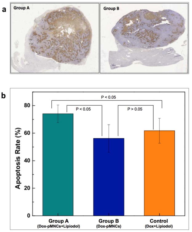
Fig. 5.
(a) Histological sections of VX2 liver tumors 14 days after treatment were TUNEL stained. The representative images from Group A and B rabbits are shown. Scattered positive cells were found study groups but a significant increase was observed for Group A (Dox-pMNCs within Lipiodol), and (b) quantitative analysis of TUNEL positive cells for Groups A, B and control. Data (means ± SD) represent the means of independent experiments (each n=5 in Group A and B, control). *p<0.05. (Group A and B). Increased TUNEL positive cells were observed in Group A vs. Group B and Group A vs. Control.

