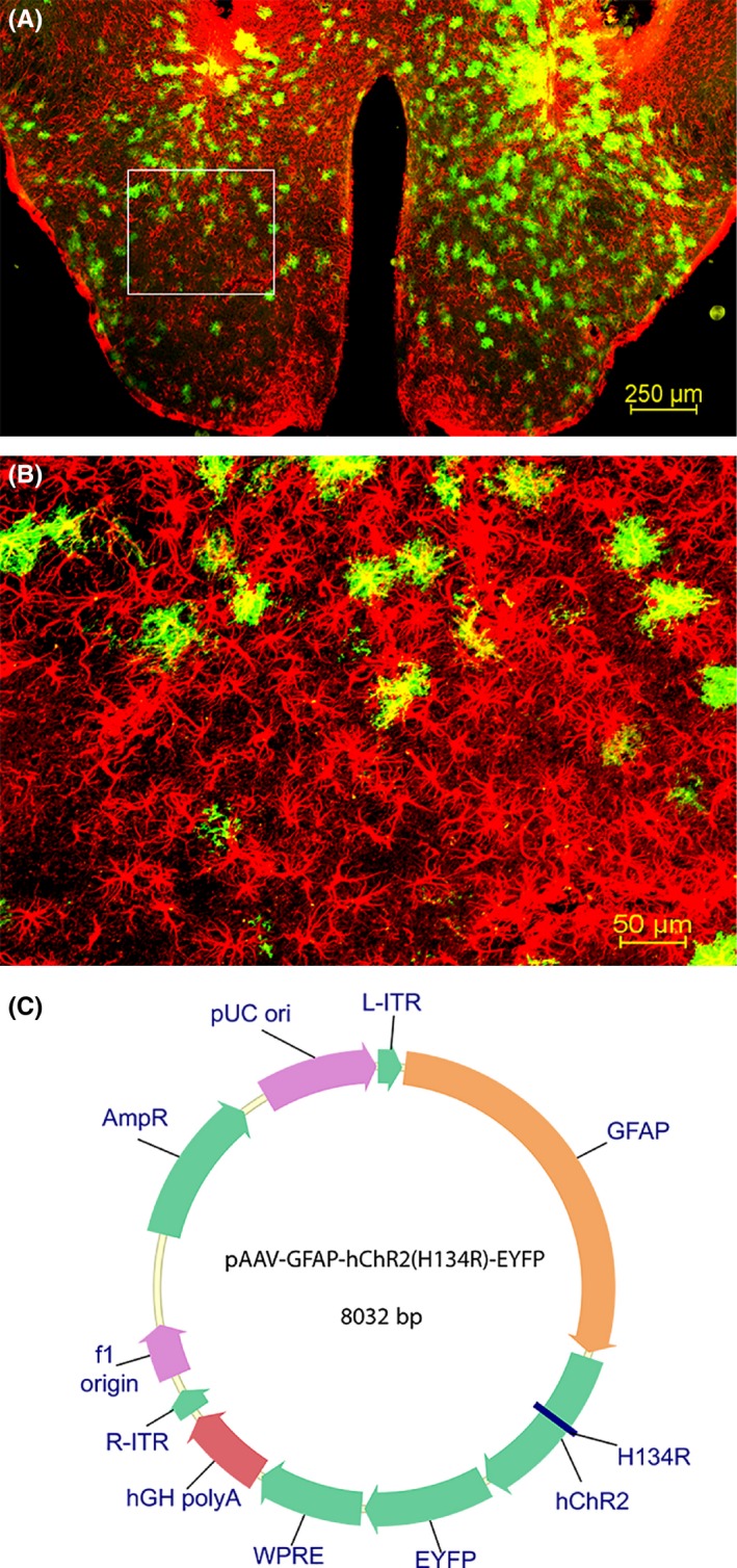
Figure 1.

Distribution of astrocytes in the posterior hypothalamus. The photomicrographs illustrate the astrocytes containing GFAP (red) and EYFP (green) in a representative mouse (A and B). A low‐magnification view of the area of disbursement of the ChR2‐EYFP in the posterior hypothalamus is depicted in A. B is a high‐magnification view of the boxed area in A. Map of ChR2 linked GFAP reporter plasmid that was packaged into recombinant adenoassociated virus (rAAV; C).
